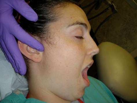
Hopefully your path to finding the solution to your problem begins here. After you have contacted our office, 301-805-9400, download and fill out the NP Forms 24.3. Most important is the Facial Problem Questionnaire. Please take you time and fill out as accurately as possible. Once complete please mail them back to us so Dr. Droter can determine what type of appointment will best suit your needs.
There are several different exams that we provide for new patients. Below are explanations of each type.
Emergency Exam
Preliminary Exam
Review of Scans
Complete Exam
Emergency Exam
A short, problem focused exam to address an immediate concern. The goal is to find a palliative solution to alleviate pain, discomfort, or worry, so that a more thorough exam can be performed at a future date. Some form of treatment or therapy may be rendered at this appointment to help alleviate pain. There will not be time at this appointment to discuss a complex history. The Facia Problem Questionnaire Form (part of New Patient Forms ) is crucial to helping Dr Droter figure out your problem efficiently and accurately, so be sure to take your time and complete the forms thouroughly.
Sample trreatment that may be recommeded: Cold Laser, Jaw exercises, Active stretching, Transcutaneous Electro Nerve stimulation (TENS), anterior stop muscle deprogrammer, triggerpoint therapy, ischemic pressure, Thermal therapy (Hot, Cold, Hot), anesthetic nerve blocks.
Preliminary Exam
Appointment Goal – To begin the process of finding the source of the pain or dysfunction. To begin to look at each area of the head and neck that may be the source of the problem. To determine whether the TMJs are healthy or damaged, and (if damaged) whether or not it is causing the problem with which the patient presents. To look for other possible causes of pain and dysfunction including cancer, neurological, vascular, muscular, and/or referred pain.

A written history is reviewed and an oral history is taken of the problem. A preliminary head and neck evaluation is performed, including: muscle coordination, muscle palpation of the head and neck, TMJ palpation, preliminary evaluation of the oral hard and soft tissues, a cancer screening exam, TMJ auscultation with a stethoscope, TMJ Doppler auscultation, range of motion analysis, TM joint loading evaluation, intra-oral occlusal analysis, and facial bone structural analysis.

The beginning of a differential diagnosis is created, with recommendations made as to the next step. This first appointment will usually take about an hour.
The goal of the first appointment is to answer these questions:
Is the TMJ damaged?
Is the Neck Damaged?
Does this damage contribute to the problems you are having?
What further tests or radiographs, if any, are needed, to develop a definitive diagnosis?
You will be informed and educated as to what your problem might be, and will be given a plan on how best to reach a definitive diagnosis. In some cases, Dr. Droter figures out what the problem is at this appointment and treatment can be initiated. However, most cases require additional information. Once we have an accurate diagnosis, a specific treatment to your specific problem can be chosen.
Review of Scans
If you were found to have TMJ damage during the first appointment, MRI and CT scans were ordered to determine what is damaged and the extend of the damage.
Appointment Goal – To interpret the MRI and CT and relate them to the clinical data gathered. To educate the patients about their disease process.
Special computer software is used to enhance the images and the data from the DICOM files sent by the radiology center. This enhanced data is analyzed and related to clinical data previously gathered. The radiology report is reviewed and correlated to my interpretation. Since the radiologist did not
have access to all the clinical data, this often limits his initial interpretation. If there are any discrepancies between my findings and the radiologist’s report, then I will consult with the radiologist to come to a consensus on the interpretation.
The patient is informed of what we see on the scans, and what this means as far as their health is concerned. Recommendations are then made to the patient as to what their next step is to start the healing process.
Either a working diagnosis or a differential diagnosis is formulated. A written summary is given to the patient.
Complete Exam
Specialized Equipment that Dr. Droter Utilizes for evaluating Joint and muscle Health
ElectroMyoGraphy (EMG)- Surface Electrodes (not the painful needle electrodes) are used to evaluation the health of the muscles. Many patients have jaw muscle that are exhausted. This measures how exhausted they are.
Joint Vibration Analysis (JVA)- CT and MRI scans show the joint not moving. The JVA is the joint in motion. A recording device that looks like a set of headphones is placed over the TMJs. The device picks up any vibrations emitted from the joints and sends them to the computer where it is recorded. A healthy joint makes no sounds. A damage joint will emit sounds. Many of the sounds can not be heard with the human ear but are detectable with this device. This information gives a picture of what happens to the joint in motion. This information is combined with the CR and MRI to give an over assessment of joint health and function.
Jaw Tracker Motion Analysis- A magnet is placed on the lower jaw. With a recording headset on, the movements of the lower jaw are tracked. If there is any joint damage or muscle disharmony. the jaw will move irregularly and be detected by this test.
TekScan Bite Analysis- A pressure sensing thin plastic is used to record the intensity and timing of which teeth hit. Ideally when a person bites down all the teeth should hit evenly. This device measures not only which teeth are hitting, but also the relative intensity and the timing.